
basic anatomy
Bones of the face and head
Cranium
The Cranium is essentially the skeleton of the head - it can be divided into the neurocranium and the viscerocranium. [6]
Neurocranium (Figure 1)
The neurocranium refers to the bones that encase and protect the brain. It can be subdivided into the Calvarium (the ‘roof’), and the Cranial Base (the ‘floor’). [6] [7]
Bones of the calvarium include: [6] [7] [8]
-
Frontal bone
-
Occipital bone
-
Parietal bones
The bones of the cranial base are comprised of the: [6] [7] [8]
-
Frontal bone
-
Occipital bone
-
Temporal bones
-
Ethmoid bones
-
Sphenoid bones
It is important to recognise that the cranium is a fixed space - any increase in mass will lead to an exponential increase in pressure. This is important when discussing the pathophysiology of traumatic brain injuries, as will be discussed in other sections. [6] [8]
Viscerocranium (Figure 2)
The viscerocranium refers to the bones of the face. Although not directly relevant to the study of traumatic brain injuries, it is helpful to have a complete understanding of the cranial anatomy. [6] [9]
-
Frontal Bone - this forms the forehead.
-
Zygomas - Commonly referred to as the “cheek bones,” these bones assist in forming the orbital cavity. They articulate with the frontal, temporal, sphenoid and maxillary bones. [6]
-
Lacrimal - These are the smallest bones of the face, and form the medial portion of the orbital cavity. [6] [9]
-
Nasal Bones - These bones are located along the bridge of the nose. [6]
-
Vomer - Posterior portion of the nasal septum. [6]
-
Palatine - These are situated at the back of the oral cavity, and form the hard palate of the oral cavity. [6] [9]
-
Maxilla - Contribute the greatest part to the upper potion of the face. Comprises of the upper jaw and along with the mandible houses teeth. [6] [9]
-
Mandible - Forms the lower, moveable jaw. [6]


Figure 1 [6]

Figure 2 [6]

Figure 3 [13]

Figure 4 [13]
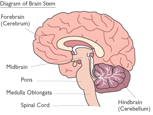
Figure 5 [18]
the brain
The brain is a magnificent organ. It is the command and coordination centre of almost every function in the body. As it is an incredibly complex organ, the intricacies won’t be fully detailed - the aim will be to refresh the basic anatomy and functions of the brain.
The brain can be divided into different regions. [10]
Cerebrum [Figure 3]
The cerebrum is comprised of two hemispheres that are separated by the falx cerebri within the longitudinal cerebral fissure. [10] [11] The cerebrum is the home to higher brain functions, and is classified in to four lobes: [10]
-
Frontal Lobe: Higher intellect, personality, mood, social conduct and language. [11]
-
Parietal Lobe: Language, calculation and visuospatial functions. [11]
-
Temporal Lobe: Memory, language, hearing. [11]
-
Occipital Lobe: Location of primary visual cortex [11]
Cerebellum [Figure 4]
Located inferiorly to the occipital lobe of the cerebrum. The cerebellum is primarily involved in coordinating movements and includes: [10] [12]
-
Planning movements [12] [13]
-
Motor learning [12]
-
Coordination of muscle activation [12] [13]
-
Error correction during movements [12]
-
Processes proprioceptive information [12]
-
Balance (receives and interprets information from the vestibular system) [12] [13]
-
Occular reflexes [13]
Brain Stem [Figure 5]
The brain stem forms the inferior portion of the brain, and connects the brain to the spinal cord via the foramen magnum. The brain stem can be divided into three different components: [14]
Medulla Oblongata
The medulla oblongata is the most inferior portion of the brain stem. It is responsible for several important functions and connects the higher levels of the brain to the spinal cord. [14] [15]
Functions include: [14] [15]
-
Basic autonomic nervous system coordination [15]
-
Respiration [14] [15]
-
Cardiac function [14] [15]
-
Vasodilation [14] [15]
-
Vomiting [14]
-
Coughing [15]
-
Sneezing [15]
-
Swallowing [14] [15]
Pons
The pons is the middle portion of the brain stem. It essentially acts as a bridge, with the following function: [14] [16]
-
Facilitates transmissions from the cerebrum to the cerebellum and medulla [14] [16]
-
Tracts that carry sensory information up into the thalamus [14] [16]
Midbrain
The midbrain is the most superior portion of the brain stem and has a variety of functions / associatioms, which include: [14] [17]
-
Eye movement [14]
-
Hearing [14] [17]
-
Motor movement [14] [17]
-
Alertness / wakefulness [14]
-
Sleep [14] [17]
-
Temperature control [14] [17]
-
Motivation [14] [17]
The meninges refer to the membranous covering of the brain and spinal cord. [19] [20] Two of the main functions include providing a framework for cerebral / cranial blood vessels, and providing a space for cerebrospinal fluid which acts to protect the brain from mechanical damage. [20] Understanding the anatomy of the meninges is important when discussing traumatic brain injuries, as many bleeds are described by their location in relation to the meninges.
There are 3 meningeal layers: [19]
Dura Mater
This is the most outermost layer, and is tightly adhered to the periosteum of the skull. [20] It is a thick, tough membrane. It can be further divided into two layers - the periosteal layer, and the meningeal layer. Between these layers lie the dural venous sinuses, which are responsible for the venous vasculature of the brain. [19] [20]
Arachnoid Mater
The middle meninge is the arachnoid mater - it is located directly underneath the dura mater. [19] [20] The space under the arachnoid mater is termed the subarachnoid space. [20] Various blood vessels run through this space. Within the subarachnoid space is cerebrospinal fluid, which assists in reducing mechanical damage to the brain by providing cushioning. [19] [20]
Pia Mater
This is the most innermost layer of the meninges. It adheres tightly to the surface of the brain, following the contours and gyri. [20] It is highly vascularised, as blood vessels travel through the membrane to perfuse the underlying brain. [19]
Meninges
the cranial nerves
Although not directly related to the pathophysiology of TBI, basic knowledge of the cranial nerves can be helpful in assessing certain neurological abnormalities following a injury. [21] Furthermore, it is compression of certain cranial nerves that can provide clues alerting us to imminent brainstem herniation.
[21] [22]